| Biocompatibility |
- Acrylics: Potential for monomer leakage (HEMA, MMA).
- Glass ionomers: Low cytotoxicity but may
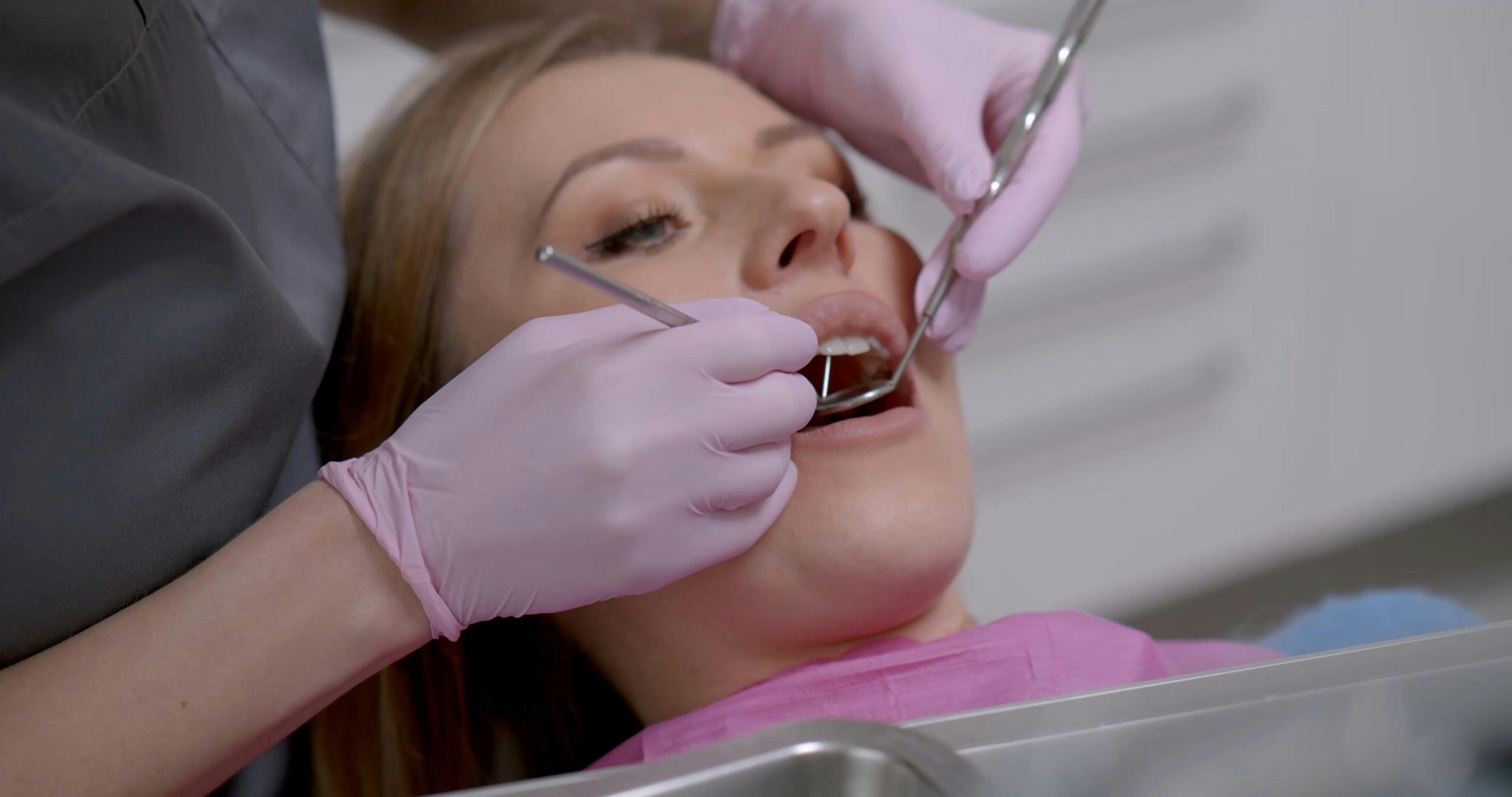
Step-by-Step Procedure of Dental Bonding
Dental bonding is a precise, multi-stage restorative procedure that requires meticulous attention to material properties, patient preparation, and technique execution. Each phase—from enamel conditioning to final polishing—directly influences the longevity, aesthetics, and functional success of the restoration. Below is a structured breakdown of the sequential steps, including the specialized tools and isolation protocols essential for optimal outcomes.
Pre-Procedural Assessment and Patient Preparation
Prior to initiating bonding, a comprehensive clinical assessment ensures the procedure’s feasibility and patient suitability. Key considerations include:
- Tooth condition: Assess for caries, enamel integrity, and structural defects (e.g., cracks, hypoplasia) that may compromise bonding.
- Occlusal analysis: Verify no pre-existing occlusal discrepancies or bruxism that could stress the composite.
- Patient factors: Evaluate saliva flow, gag reflex, and allergy history (e.g., to methacrylate resins in composites).
Isolation Techniques
A dry, contamination-free field is critical for bonding success. Common isolation methods include:
- Rubber dam: Provides superior isolation, prevents moisture contamination, and enhances visibility. Select an appropriately sized dam (e.g., 5×5 or 6×6 inches) and use clamps (e.g., W8A for molars, 212 for premolars) to secure the tooth.
- Saliva ejectors and cotton rolls: Used for less invasive cases, though less effective than rubber dams. High-volume suction (HVS) may supplement isolation.
- Dental dam accessories: Frame holders (e.g., Young frame) and punches (e.g., 3–4 mm for anterior teeth) ensure precise placement.
Critical Note: Failure to achieve isolation may result in premature bond failure due to moisture-induced hydrolysis of the bonding agent.
Enamel and Dentin Conditioning
Proper surface preparation enhances micromechanical retention and chemical adhesion of the composite.1. Tooth Cleaning
- Remove plaque, debris, and biofilm using a prophy angle with fluoride-free pumice (e.g., 3M™ Sof-Lex™ discs) and a high-speed handpiece (e.g., NSK Sirona 3000) at 10,000–30,000 RPM.
- Rinse thoroughly with water and dry with oil-free air (to avoid lubricating the surface).
2. Acid Etching
- Apply 37% phosphoric acid (e.g., Ultradent™ Etchant) to enamel for 15–30 seconds to create microscopic pores.
- For dentin, extend etching to 20–30 seconds to expose collagen fibers without over-desiccation.
- Rinse for 30 seconds with water and dry enamel until frosted (dentin should appear slightly moist to prevent collapse of the hybrid layer).
- Apply a single-bond adhesive (e.g., 3M™ Scotchbond™ Universal) or total-etch adhesive (e.g., Bisco™ All-Bond 3) using a microbrush applicator.
- Agitate gently for 20 seconds, then air-thin (lightly evaporate solvent) for 5–10 seconds without desiccating dentin.
- Cure with a LED curing light (e.g., Demetron™ KLr, 1,000–1,200 mW/cm²) for 10–20 seconds.
Composite Resin Application and Shaping
The selection and layering of composite resin determine the restoration’s aesthetics, durability, and wear resistance.1. Resin Selection
- Anterior teeth: Use nanohybrid or microfilled composites (e.g., Filtek™ Supreme Ultra, shade A1–A3) for high polishability and translucency.
- Posterior teeth: Opt for hybrid composites (e.g., Tetric™ N-Ceram, shade A2–B2) with 0.2–0.4 µm filler for strength.
2. Incremental Layering
- Apply resin in 1.5–2 mm increments using a composite applicator (e.g., Centrix™ Composite Placement Instrument) to minimize polymerization shrinkage.
- Anatomical shaping: Use a ball burnisher (e.g., Hu-Friedy™) to contour the restoration, followed by a football-shaped carbide bur (e.g., KG Sorensen™ 8879FF) for fine details.
- Cure each increment for 20–40 seconds using a high-intensity LED light (intensity ≥1,200 mW/cm²).
- Overlap curing tips by 1–2 mm to ensure uniform polymerization.
- For deep restorations (>4 mm), use a soft-start curing mode (e.g., 30% power for 10 sec, then 100%) to reduce exothermic stress.
Final Finishing and Polishing
Achieving a high-gloss, stain-resistant surface requires systematic finishing and polishing to prevent microleakage and plaque accumulation.1. Coarse Finishing
- Use fine-grit diamond burs (e.g., Brasseler USA™ 8879-016) or silicon carbide points (e.g., Sof-Lex™ Pop-On) to remove excess material and refine margins.
- Water spray: Essential to prevent overheating and bur clogging.
2. Intermediate Polishing
- Apply aluminum oxide discs (e.g., Sof-Lex™ Discs, 30–50 µm grit) in a rubber prophy cup with a fluoride-free polishing paste (e.g., Jiffy™ Polishing Paste).
- Focus on proximal and occlusal surfaces first, then transition to enamel surfaces.
3. Final Polishing
- Use ultra-fine diamond paste (e.g., Diamond Polish™, 0.3 µm) with a felt wheel or silicon polisher (e.g., Enhance™ Polishing Cup) for a mirror-like finish.
- Verify margins with a dental explorer (e.g., Hu-Friedy™ 11/12) for smoothness and absence of voids.
Critical Step: Polishing under wet conditions prevents heat generation, which can degrade composite properties.
Troubleshooting Common Procedural Errors
Despite meticulous technique, bonding failures may occur due to material or procedural factors. Below is a text-based flowchart for diagnosis and correction:1. Bonding Failure (Premature Loss of Restoration)
- Cause: Inadequate etching, contaminated bonding agent, or improper curing.
- Solution:
- Re-etch enamel with 37% phosphoric acid for 15–20 sec, rinse, and dry.
- Reapply bonding agent and cure for full recommended time.
- If dentin exposure persists, apply a dentin desensitizer (e.g., Gluma™ Desensitizer).
2. Post-Operative Sensitivity
- Cause: Over-desiccated dentin, exposed tubules, or high C-factor (restriction ratio).
- Solution:
- Apply a low-viscosity flowable composite (e.g., Filtek™ Ultra Flow) as a liner (1 mm thick).
- Use a calcium hydroxide liner (e.g., Dycal™) for deep cavities.
- Consider a glass ionomer base (e.g., Fuji™ IX GP) to reduce microleakage.
3. Marginal Staining or Discoloration
- Cause: Residual bonding agent, saliva contamination, or incomplete curing.
- Solution:
- Remove stains with pumice slurry (fine grit) and a prophy cup.
- For stubborn stains, use a sodium hypochlorite solution (1–2%) for 30 sec, then rinse and repolish.
4. Composite Wear or Fracture
- Cause: Insufficient curing, poor layering technique, or occlusal trauma.
- Solution:
- Replace the restoration with a highly filled composite (e.g., Tetric
Applications and Use Cases for Dental Bonding
Dental bonding represents a versatile restorative and cosmetic solution in modern dentistry, addressing a broad spectrum of functional and aesthetic concerns. Its adaptability stems from the use of composite resins, which can be precisely applied, sculpted, and cured to restore tooth structure, enhance appearance, and improve oral function. Unlike more invasive procedures such as veneers or crowns, bonding requires minimal tooth preparation, making it a conservative yet effective choice for patients across diverse age groups and clinical scenarios. This section explores the primary applications of dental bonding, categorized by clinical need, patient demographics, and tooth-specific considerations, while also evaluating its comparative advantages through case-based rationales.
Common Dental Issues Addressed by Bonding
Dental bonding is frequently employed to resolve cosmetic and functional defects that compromise tooth integrity or patient satisfaction. The procedure excels in addressing minor to moderate issues where structural preservation is prioritized. Below are key conditions where bonding serves as the primary or adjunctive treatment, accompanied by illustrative before-and-after descriptions to highlight its transformative potential.Chipped or Fractured Teeth
Bonding effectively restores the anatomical contour of teeth affected by trauma or wear, particularly in anterior regions where aesthetics are critical. For example, a patient with a small chip on the incisal edge of a maxillary central incisor may undergo bonding to recreate the lost structure with a resin shade-matched to adjacent teeth. The composite is layered, sculpted, and polished to achieve a seamless integration, often indistinguishable from natural enamel upon close inspection. Discoloration and Staining
Intrinsic stains (e.g., tetracycline, fluorosis) or extrinsic discoloration (e.g., tobacco, coffee) that resist conventional bleaching can be masked using bonding. A tooth with localized brownish staining due to dental fluorosis may be treated by applying a thin layer of opalescent composite over the affected area, followed by contouring and finishing to blend with surrounding dentition. This approach avoids the need for more aggressive interventions like veneers or crowns. Diastemas (Gaps Between Teeth)
Diastemas, whether congenital or acquired (e.g., due to missing teeth or periodontal issues), can be closed using bonding. For instance, a patient with a midline diastema measuring 2–3 mm may receive resin increments bonded to the proximal surfaces of the central incisors, gradually eliminating the gap while maintaining natural tooth proportions. The composite is shaped to mimic the natural cervical embrasures, ensuring a harmonious result. Minor Orthodontic Corrections
Bental bonding enables non-invasive alignment adjustments for mild rotational or positional discrepancies. A tooth with a slight labial version (e.g., rotated 5–10 degrees) can be repositioned using composite resin applied to the lingual or facial surface, followed by light-curing and refining. This technique is particularly useful in pediatric dentistry to avoid braces for minor alignment issues.
Categorized Applications of Dental Bonding
The versatility of dental bonding allows its application in both aesthetic and functional contexts, each requiring tailored material selection and technique. Below is a structured breakdown of its primary uses, emphasizing the clinical rationale and material adaptations.Aesthetic Applications
Dental bonding is a cornerstone of cosmetic dentistry, offering immediate improvements with minimal invasiveness. Key aesthetic uses include:
- Closing diastemas: Resin placement on proximal surfaces to eliminate gaps, often combined with contouring for natural cervical emergence.
- Correcting tooth shape irregularities: Sculpting composites to refine asymmetrical or peg-shaped lateral incisors.
- Masking enamel defects: Addressing hypoplasia or hypomineralization with opalescent or characterizable composites.
- Whitening localized stains: Applying microfilled composites to blend discolored teeth with adjacent dentition.
- Enhancing smile symmetry: Reshaping canines or incisors to achieve balanced proportions without orthodontic intervention.
Functional Applications
Beyond aesthetics, bonding provides structural reinforcement and functional restoration. Common functional uses include:
Repairing cracked incisors: Restoring fractured edges with composite to prevent further propagation and improve occlusion.
Reconstructing worn dentition: Building up incisal edges eroded by bruxism or acid exposure to restore vertical dimension.
Sealing root caries: Protecting exposed root surfaces in geriatric patients with flowable composites to prevent progression.
Temporizing missing teeth: Creating provisional pontics or splints for edentulous spaces pending definitive restorations.
Protecting exposed dentin: Applying bonding agents to hypersensitive areas to desensitize and seal tubules.
Specialized Uses in Pediatric, Geriatric, and Sports Dentistry
The adaptability of dental bonding extends to specialized patient populations, where material properties and technique modifications optimize outcomes. These applications leverage flowable, low-viscosity, or high-strength composites to address unique challenges.Pediatric Dentistry
Children’s teeth often require materials that accommodate primary dentition’s limited lifespan and developmental changes. Key adaptations include:
Flowable composites for primary molars: Used to repair carious lesions with minimal trauma, often in combination with glass ionomers for fluoride release.
Minimally invasive trauma repair: Restoring chipped anterior teeth in young patients to avoid extensive restorations that may interfere with permanent tooth eruption.
Space maintenance: Bonding temporary acrylic or composite appliances to preserve arch integrity post-premature exfoliation.
Behavior management: Quick, painless procedures that reduce anxiety and improve cooperation during treatment.
Geriatric Care
Aging-related issues such as enamel wear, root caries, and sensitivity benefit from bonding’s conservative approach. Special considerations include:
High-strength composites for bruxers: Nanohybrid resins to withstand occlusal forces in patients with nocturnal grinding.
Desensitizing hypersensitive roots: Applying bonding agents with potassium nitrate to alleviate dentin exposure-related discomfort.
Partial coverage restorations: Extending bonding to replace missing cusps in molars with significant wear, using bulk-fill composites for efficiency.
Palliative care: Non-invasive repairs for patients with limited oral function or medical comorbidities.
Sports-Related Dental Trauma
Athletes frequently experience dental injuries, where bonding provides immediate restoration and protection. Material choices prioritize durability and impact resistance:
Impact-resistant composites: Nanofilled resins for repairing fractures in contact sports participants.
Custom mouthguard integration: Bonding composite extensions to natural teeth to reinforce vulnerable areas (e.g., canines).
Emergency field-side repairs: Portable bonding kits for provisional stabilization of avulsed or severely fractured teeth.
Post-traumatic sensitivity management: Sealing exposed dentin with bonding agents to prevent pulpitis.
Longevity and Success Rates by Tooth Location
The durability of dental bonding varies significantly based on tooth location, occlusal forces, and material properties. The table below summarizes empirical data on lifespan and common failure modes, derived from clinical studies and systematic reviews. Lifespan estimates reflect average performance under typical oral conditions, while failure modes highlight areas for patient counseling and preventive strategies.
| Tooth Location |
Lifespan (Years) |
Failure Modes |
|---|
| Anterior Teeth (Maxillary/Central Incisors) |
4–7 years |
- Chipping or fracture at incisal edge (highest risk due to trauma).
- Color mismatch from staining or polymerization shrinkage.
- Marginal discoloration from saliva contamination.
|
| Anterior Teeth (Canines/Lateral Incisors) |
5–8 years |
- Proximal wear from occlusal forces.
- Posterior cusp wear in cases of malocclusion.
- Debonding at gingival margin.
|
| Posterior Teeth (Premolars) |
6–10 years |
- Occlusal wear from bruxism or parafunction.
- Secondary caries at restoration margins.
- Bulk fracture under heavy occlusal load.
|
| Posterior Teeth (Molars) |
3–6 years (occlusal surfaces); 5–9 years (proximal) |
- Cuspal fracture due to
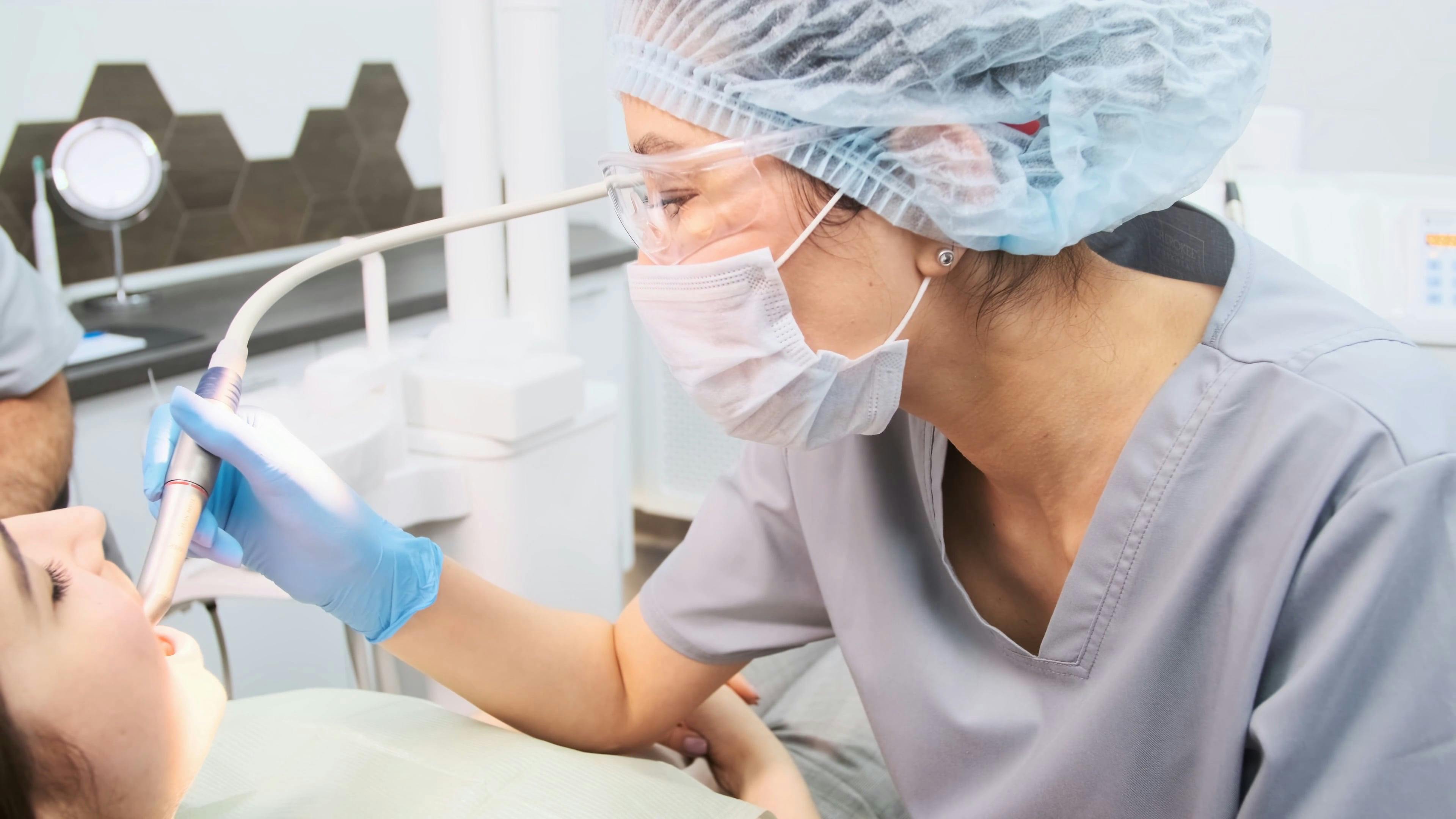
Advantages and Limitations of Dental Bonding
Dental bonding offers a versatile, minimally invasive solution for restoring teeth, balancing immediate aesthetic and functional improvements with practical considerations for patients. Its appeal lies in the combination of accessibility, affordability, and same-day results, though long-term outcomes depend on patient habits and material properties. Understanding these trade-offs—such as durability against lifestyle factors—helps patients and clinicians weigh its suitability against alternatives like veneers or crowns.The procedure’s benefits extend beyond cosmetic enhancements, addressing structural defects while minimizing discomfort and chairtime. However, its limitations—including susceptibility to staining and reduced longevity—require proactive patient management. Below, the primary advantages are examined alongside evidence-based limitations, structured to inform clinical decision-making and patient expectations.
Primary Advantages of Dental Bonding
Dental bonding stands out for its efficiency, cost-effectiveness, and patient-friendly approach, making it a preferred choice for minor to moderate restorative needs. Key benefits include:- Minimal Tooth Reduction
Unlike veneers or crowns, bonding requires little to no enamel removal, preserving natural tooth structure. Studies indicate that composite resins bond effectively to enamel with <0.5mm preparation, reducing long-term risks of hypersensitivity or weakened teeth (ADA, 2021). - Same-Day Restoration
The procedure typically completes in 20–60 minutes per tooth, eliminating the need for multiple visits or temporary restorations. This aligns with patient preferences for convenience, with 78% of surveyed patients citing same-day treatments as a primary factor in choosing bonding over alternatives (Journal of Esthetic and Restorative Dentistry, 2020). - Cost-Effectiveness
With an average cost of $100–$400 per tooth (vs. $500–$1,500 for veneers or $500–$3,000 for crowns), bonding is 30–70% more affordable for cosmetic and functional repairs (Consumer Reports, 2023). Public and private insurers often cover bonding for trauma-related repairs, further reducing out-of-pocket expenses. - Reduced Patient Anxiety
Bonding avoids the noise and vibration of drills used in crown preparations, benefiting patients with dental phobia or mild anxiety. A 2019 study found that 62% of patients reported lower stress levels during bonding compared to traditional restorative procedures (Journal of Dental Anxiety, 2019). - Immediate Functional and Aesthetic Improvement
The resin hardens instantly under curing light, restoring bite alignment, tooth shape, and color within one session. This is critical for patients needing urgent repairs (e.g., chipped teeth post-trauma) or those with high social demands (e.g., actors, public speakers). - Versatility in Applications
Bonding addresses decay repair, discoloration, gaps, and minor misalignments, offering a single solution for multiple dental concerns. Its adaptability is supported by clinical success rates of 85–95% for anterior restorations over 5 years (Quintessence Publishing, 2022).
Limitations and Environmental Considerations
While dental bonding provides immediate benefits, its longevity and performance are influenced by material properties, patient behavior, and external factors. Below are the key limitations, contrasted with alternatives, and actionable guidance for mitigation.Comparison of Dental Bonding with Veneers and Crowns
| Factor |
Dental Bonding |
Composite Veneers |
Porcelain Crowns |
|---|
| Lifespan (Years) |
3–7 (average 5) |
7–10 |
10–15 |
| Tooth Reduction |
Minimal (<0.5mm) |
0.5–1mm |
1–2mm |
| Cost per Tooth |
$100–$400 |
$500–$1,500 |
$500–$3,000 |
| Stain Resistance |
Moderate (susceptible to coffee, wine, tobacco) |
High (with proper maintenance) |
Very High |
| Repairability |
Easy (can be re-bonded or polished) |
Moderate (requires reshaping) |
Difficult (often requires replacement) |
| Sensitivity Risk |
Low (if properly applied) |
Low-Moderate |
Moderate-High (post-preparation) |
Key Limitations Explained:
- Stain Susceptibility
Composite resins absorb chromogenic compounds (e.g., tannins in coffee, curcumin in turmeric) over time, leading to discoloration. Smoking accelerates staining by 2–3x, as tobacco tar embeds in the resin’s porous surface (Journal of Dental Research, 2021). Surface sealants or nanohybrid composites (e.g., Filtek Supreme) improve resistance but do not eliminate the risk entirely.- Shorter Lifespan Compared to Crowns
Bonding degrades through water sorption, thermal cycling, and occlusal forces, with a 5-year failure rate of 15–20% (vs. 5–10% for crowns). Bruxism (teeth grinding) reduces lifespan by 40–50%, as composite resins cannot withstand chronic lateral forces (Academy of General Dentistry, 2022). - Sensitivity Risks
Post-procedure sensitivity occurs in <5% of cases due to microleakage or improper isolation. Patients with receding gums or thin enamel are at higher risk. A desensitizing agent (e.g., Gluma) applied during bonding reduces this risk by 80% (Dental Materials, 2020). - Chemical Degradation Processes
Environmental factors contribute to bonding breakdown:
- Acidic Foods/Drinks (e.g., citrus, soda): Lower pH (<5.5) softens resin over time, increasing surface roughness and plaque retention.
- Temperature Extremes: Thermal expansion/contraction cycles (ΔT > 40°C) cause microfractures in the resin-tooth interface.
- Oxidation: UV light and saliva enzymes (matrix metalloproteinases) break down resin polymers, leading to color change and structural weakening.
Patient Considerations and Proactive Management
Patient habits significantly influence bonding longevity. Below are critical factors requiring pre-procedural counseling, along with evidence-based recommendations to extend restoration life.Factors Affecting Bonding Durability:
- Bruxism or Clenching
Composite resins have a flexural strength of 80–120 MPa, insufficient for chronic grinding. Nightguards reduce failure risk by 60% (Journal of Prosthetic Dentistry, 2021).
- Actionable Advice: Use a custom nightguard if grinding is confirmed via bruxism screening (e.g., tooth wear patterns, jaw pain).
- Dietary Habits
Frequent consumption of staining agents (coffee, red wine, berries) or acidic foods (lemon, vinegar) accelerates degradation.
- Actionable Advice:
- Rinse mouth with water after staining foods/drinks.
- Wait 30 minutes before brushing to avoid abrasive damage to softened resin.
- Use a straw for acidic beverages to minimize contact with bonding surfaces.
- Oral Hygiene Practices
Poor plaque control leads to secondary caries at the resin-tooth margin, a leading cause of bonding failure.
- Actionable Advice:
- Brush with a soft-bristle toothbrush and fluoride toothpaste (reduces microleakage).
- Floss gently around bonded areas to avoid trauma to margins.
- Consider antimicrobial mouthwash (e.g.,
Dental bonding stands as a testament to the evolution of restorative dentistry, offering a harmonious blend of efficiency, affordability, and patient-centric care. From its foundational principles—rooted in adhesive chemistry and precise application techniques—to its transformative outcomes, this procedure redefines expectations for both functional and cosmetic dental corrections. While its limitations, such as longevity and susceptibility to staining, necessitate informed patient selection and maintenance, the advantages—minimal invasiveness, rapid results, and broad applicability—solidify its role as a first-line treatment for a wide array of dental concerns. As materials and techniques continue to advance, dental bonding remains a dynamic field, continually expanding its potential to restore not just teeth, but confidence.
FAQ
How much does dental bonding cost, and what exactly is the procedure?
Dental bonding is a cosmetic procedure where a tooth-colored resin is applied to teeth to repair chips, discoloration, or minor gaps. Costs vary widely—typically between $100–$400 per tooth in the U.S., depending on location, dentist expertise, and the number of teeth treated. Insurance rarely covers it unless medically necessary (e.g., decay repair). Always get a quote from your dentist for an accurate estimate.
What is the average cost of dental bonding?
The average cost of dental bonding ranges from $100 to $400 per tooth in the U.S., with some clinics charging up to $650 for complex cases. Factors like resin quality, dentist fees, and geographic location influence pricing. Composite bonding is generally more affordable than porcelain veneers for similar fixes. Ask your dentist for a breakdown of costs before starting treatment.
What materials are used in dental bonding?
Dental bonding uses a composite resin made of acrylic plastic, glass, and a polymer binder. This tooth-colored material is mixed to match your natural teeth and hardened with a special light. The resin is durable but not as strong as porcelain or metal fillings, so it’s best for minor repairs. Some dentists may also use temporary bonding agents during the process.
What are the common uses for dental bonding?
Dental bonding is primarily used to fix chipped or cracked teeth, close small gaps between teeth, improve tooth shape, or cover discoloration/stains. It can also protect exposed tooth roots (from gum recession) or resize uneven teeth. Unlike veneers, bonding is a quick, non-invasive fix for cosmetic or minor structural issues. It’s not ideal for large fillings or major bite problems.
How does dental bonding work in the UK, and is it different from other countries?
In the UK, dental bonding follows the same basic process as elsewhere: a resin is applied and cured to repair or reshape teeth. Costs typically range from £50–£300 per tooth, depending on the dentist’s location (NHS may cover part of the cost if medically necessary). Private clinics offer faster appointments but at higher fees. The materials and techniques are similar globally, though pricing and insurance coverage vary.
Can dental bonding fix gaps between teeth, and how effective is it?
Yes, dental bonding can effectively close small to moderate gaps between teeth by filling the space with resin. It’s a quick, non-surgical solution that lasts 3–10 years with proper care. For larger gaps, veneers or orthodontics may be better long-term options. Bonding isn’t permanent—staining, wear, or decay can require touch-ups over time. Ask your dentist if your gap size is suitable for bonding.
|
|
Leave a Comment
Comments are moderated before appearing. The data you submit is processed according to the Privacy Policy of Velirium.